Osteocyte lacunae features in different chicken bones
Istituto Zooprofilattico Sperimentale del Piemonte, Liguria e Valle d’Aosta. Area Territoriale/Sezione di Aosta. CERMAS (National Reference Centre for Wildlife Diseases). Regione Amerique 7/G. I-11020 Quart (Italy). E-mail: lorenzo.domenis@izsto.it
Istituto Zooprofilattico Sperimentale del Piemonte, Liguria e Valle d’Aosta. National Reference Centre for the Surveillance and Monitoring of Animal Feed/National Reference Laboratory for Animal Proteins in Feeding Stuffs (C.Re.A.A/NRL). Via Bologna, 148. I-10154 Torino (Italy).
Istituto Zooprofilattico Sperimentale del Piemonte, Liguria e Valle d’Aosta. National Reference Centre for the Surveillance and Monitoring of Animal Feed/National Reference Laboratory for Animal Proteins in Feeding Stuffs (C.Re.A.A/NRL). Via Bologna, 148. I-10154 Torino (Italy).
Istituto Zooprofilattico Sperimentale del Piemonte, Liguria e Valle d’Aosta. National Reference Centre for the Surveillance and Monitoring of Animal Feed/National Reference Laboratory for Animal Proteins in Feeding Stuffs (C.Re.A.A/NRL). Via Bologna, 148. I-10154 Torino (Italy).
Abstract
Directive 2003/126/EC defines the method for the determination of constituents of animal origin for official control of feedingstuffs. One of the hardest problems for microscopist is the differentiation between mammalian and poultry bones on the basis of some characteristics as colour and borders of the fragments, shape and density of osteocyte lacunae. The shape of osteocyte lacuna in poultry and mammals is often described in different way, elliptic or roundish according with the Author(s). The aim of this study was to analyze the characteristics of lacunae in chicken bones of different type. For this purpose, smashed fragments and histological sections of the same bone were compared in order to evaluate the microscopic aspect of lacunae in different breaking and trimming planes. According to the observations carried out, it was possible to infer that chicken osteocyte has a biconvex lens shape; however the different arrangement and some size variation of the osteocytes in the several bone segments influence the microscopic features of corresponding lacunae. Chicken bone is made of a parallel-fibered tissue, without osteons. This structure probably determines the plane fracture of the bone and consequently the different aspect of lacunae (from spindle-shaped to elliptic-roundish) we can see in chicken derived PAP (processed animal protein). For example, in the fragments obtained from smashed diaphysis, the prevalence of spindle-shaped lacunae is depending on the preferential breaking of the bone along longitudinal plane. Likewise, for the epiphysis, being made mostly by bone trabeculae with strange directions, the breaking happens along different planes, creating lacunae of various shape. Performing the official check of animal feedingstuffs, the presence of bone fragments with roundish or elliptic osteocyte lacunae induces the analyst to thinking that the meat and bone meal comes respectively from mammals and poultry or vice versa depending to the reference Author(s); apart from the final evaluation (also based on some other features of the fragments), it is important to consider that the chicken bones could show lacunae of different shapes (spindle, elliptic and roundish), in accordance with the type and the breaking of the involved skeleton segments.
1. Introduction
1In order to avoid a further spread of BSE and its human form nvCJD, the European Commission has taken several actions. The Commission Decision 94/381/EC of 27 June 1994, amended several times, stated for the first time the prohibition of the use of proteins derived from all mammalian tissues in feedingstuffs intended for ruminants. Harmonized and more complete EU control rules introduced in 2001 included a ban, except for fish proteins under specific circumstances, on the feeding of PAP (processed animal proteins) to animals which are kept, fattened or bred for the production of food (Regulation 999/2001/EC). These control actions combined with the lab check of nervous tissue of the slaughtered ruminants, have proved successful and have significantly reduced the number of BSE cases across EU member states. Directive 2003/126/EC defines the method for the determination of constituents of animal origin for official control of feedingstuffs by microscopy. The detection limit of the provided microscopic method is approximately 0.1% or even smaller. Even though skilled and trained microscopists are involved in this activity, it is often difficult to differentiate between mammalian and poultry bones, which is done examining several characteristics such as colour and borders of the bone fragments, shape and density of osteocyte lacunae. Some of these features are not defined with one accord through the scientific community; especially the shape of osteocyte lacuna is described by some Authors as elliptic in poultry and roundish in mammals (Gasparini et al., 1996), while in other works (Gizzi et al., 2003) poultry lacunae are defined more globular compared to those of mammals. Since the examination is visual, the species definition often depends only on the microscopist’s experience, although recently it has been proposed the application of image analysis for some parameters of bone lacuna (Pinotti et al., 2004). The aim of this study was to give a precise description of the osteocyte lacuna morphology in chicken bones of different type, useful for distinguishing among land animals in PAP identification and characterization.
2. Material and methods
2For this purpose a 60 days old broiler chicken baked at 220°C was used. After the stripping of flesh, different types of bones were examined, as long bones (thigh bone, shinbone, homer, radius and ulna, metacarpus, metatarsus), short bones (vertebrae) and flat bones (rib, sternum, shoulder bone and skull).
3The selected bone portions (diaphysis and epiphysis of long bones, different segments of vertebrae and flat bones) were partly fixed with formaldehyde 10% for the histological analysis, partly smashed with a pestle for the direct microscopic examination.
4Samples for the histological exam were decalcified for one or two days (depending on their own size) with Osteodec (Carlo Erba), trimmed upon longitudinal, cross and tangential sections and then dehydrated with increasing concentrated alcohol solutions. Afterwards, they were cleared with Bioclear (Byoptica) and embedded in paraffin wax for sectioning on microtome. Slices of 5 micrometers thickness were stained with Haematoxylin Eosin (HE) method, mounted with cover glass using Eukitt balsam and observed in bright field at different magnifications with Olympus BX 60 compound microscope, equipped with Digital Camera C40/40 Zoom and software Olympus DPSoft (Version 3.1) for microphotograph.
5Direct microscopic examination on smashed bones was carried out with the same instruments, previously embedding the fragments in fixing oil.
3. Results
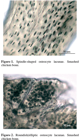
6It was possible to evaluate the microscopic aspect of osteocyte lacunae in different trimming planes by comparing smashed fragments and histological sections of the same bone portion. Generally, in smashed diaphysis of long bones the lacunae appeared mostly spindle-shaped (Figure 1), with a lower presence of roundish-elliptic type (Figure 2).

7There was not a prevalent shape in smashed long bones epiphysis, flat bones, vertebrae where it was possible to find spindle-shaped, roundish or elliptical lacunae indifferently.
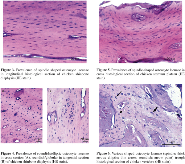
8For histological sections, the trimming was made according to different planes of the space. Referring to diaphysis’portions, in longitudinal section the lacunae appeared mostly spindle-shaped (Figure 3), in cross section elliptic-roundish, in tangential section mostly globular (Figure 4).
9In cross section, the flat bones showed mostly spindle shaped lacunae in the plateaux (Figure 5), while in the trabecular inner portion, as in the epiphysis of long bones and vertebrae (Figure 6), the lacunae could appear of various shape with different prevalence (spindle, elliptic or roundish).
4. Discussion and conclusion
10According with the observations carried out in the different planes of trimming, we infer that chicken osteocyte, as it happens in the man (Bani, 2007), has a biconvex lens shape. This morphology seems to be maintained in all skeleton parts; however the different arrangement and some size variation of the osteocytes in the various bone segments influence the microscopic features of corresponding lacunae. Chicken bone is made of a parallel-fibered tissue, without osteons (Barasa, 1983). This distinctive structure probably determines the plane fracture of the bones and consequently the different aspects of lacunae (from spindle-shaped to elliptic-roundish) we can see in chicken derived PAP. For example, in the fragments from the smashed diaphysis, the prevalence of spindle-shaped lacunae depends on the preferential breaking of the bone along longitudinal plane. Likewise, for the epiphysis, being made (besides of cartilage) mostly of bone trabeculae with strange directions, the breaking happens along different planes, creating lacunae of various aspect. Performing the official check of animal feedingstuffs, the presence of bone fragments with roundish or elliptic osteocyte lacunae induces the analyst to thinking that the meat and bone meal comes respectively from mammals and poultry or vice versa depending to the reference Author(s); apart from the final evaluation (also based on some other features of the fragments), it is important to consider that the chicken bones could show lacunae of different shapes (spindle, elliptic and roundish), in accordance with the type and the breaking of the involved skeleton segments.
11Acknowledgements
12We thank E. Pepe, R. Spedicato and P. Pecoraro of the IZSPLV Section of Aosta for the histological technical support.
Bibliographie
Bani D. Il tessuto osseo. Http: www.med.unifi.it/didonline/anno-I/istologia/osso/osso.htm, (03.09.2007)
Barasa A., 1983. Istologia generale e speciale II°. Tessuti. Anno Accademico 1983-84. Torino, Italy: Cooperativa Libraria Universitaria.
Commission Decision 94/381/EC concerning certain protection measures with regard to bovine spongiform encephalopathy and the feeding of mammalian derived protein. Off. J. Eur. Union, L172, 27.06.94, 23.
Commission Directive 2003/126/EC on the analytical method for the determination of constituents of animal origin for the official control of feedingstuffs. Off. J. Eur. Union, L339, 24.12.03, 78-84.
Gasparini G. & Grisafulli A., 1996. Identificazione delle farine di carne di mammiferi nei mangimi. Tecnica Molitoria, Agosto, 766-778.
Gizzi G. et al., 2003. An overview of tests for animal tissues in feeds applied in response to public health concerns regarding bovine spongiform encephalopathy. Rev. Sci. Tech. Off. Int. Epizooti., 22(1), 311-331.
Pinotti L. et al., 2004. Microscopic method in processed animal proteins identification in feed: applications of image analysis. Biotechnol. Agron. Soc. Environ., 8(4), 249-251.
Regulation (EC) n°999/2001 laying down rules for the prevention, control and eradication of certain transmissible spongiform encephalopathies. Off. J. Eur. Communities, L147, 31.05.2001, 1-40.


